
Personalised service by a NHS Senior Ultrasonographer Scanning Privately on Weekends!
A full Consultation with your Ultrasonographer is included in the below price.
We will give you a report of your findings, but the images will only be sent to your NHS GP or Medical Clinic (with your consent).
| Diagnostic Test | Price |
|---|---|
| Breast Ultrasound (Unilateral) | £160 |
| Breast Ultrasound (Bilateral) | £200 |
- PLEASE NOTE – We do not do normal full breast health check screenings as our ultrasonographer is not trained in this. Further, we do not offer pre-surgery scanning requested for by specialists – you will need to see a breast consultant for this.
- Please arrive at least 10 minutes before your appointment time – this allows adequate time to get registered with us.
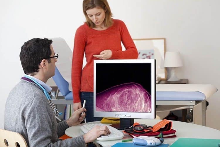
What is a Breast Ultrasound?
This is the examination of the breast tissue using an ultrasound scan. Ultrasound uses high frequency sound waves to produce images of the breast that are displayed on a screen.
Why would I need a Breast Ultrasound?
If you or your doctor can feel a lump in the breast, ultrasound can help to distinguish fluid-filled lumps (cysts) from solid lumps that may be cancerous or benign (non-cancerous). The scan evaluates a palpable lump on the breast for suspicious features and then graded BI-RADS. This is not a screening study in place of a mammogram. It can also evaluate implants for signs of rupture, but is not a pre-surgery screening ultrasound.
In younger patients who have breast symptoms (e.g. pain, lumps), ultrasound is often the first investigation. The breast tissue of younger women is much denser than it is in older women, and this can make it harder to detect an abnormality using an X-ray (mammogram).
Ultrasound is also used to diagnose problems such as complications from mastitis (an infection that occurs most often during breast-feeding), assessing abnormal nipple discharge or problems with breast implants.
If an abnormality is found during the ultrasound, you may need to be referred to the hospital to have an ultrasound-guided needle biopsy (Breast FNA or Core Biopsy).
How do I prepare for a Breast Ultrasound?
No preparation is necessary. It is advisable to wear a two-piece outfit so that only your top has to be removed to provide access to the breast area.
What happens during a Breast Ultrasound?
- You will be asked to remove your top and bra, and change into a gown.
- You will be asked to lie on a bed and one breast at a time will be examined.
- The ultrasound probe (called a transducer) is placed on the breast and gently moved around the breast to examine the breast tissue.
- Examination of the armpit (or axilla) will also be undertaken to assess for any enlarged lymph glands (or nodes), a lump or swelling.
What are the risks of a Breast Ultrasound?
There are no risks from an ultrasound. Even if you are pregnant, you are able to safely have an ultrasound examination.
What are the benefits of a Breast Ultrasound?
Ultrasound examination allows the detection and identification of most breast lumps. It is especially useful in distinguishing between solid and fluid-filled lumps.
If the ultrasound does not identify a lump that you or your doctor can feel, then other tests, such as a mammogram or magnetic resonance imaging (MRI), may be required to examine the breast.
When is a mammogram more useful?
Mammograms are used in screening in women aged 50 and over. A mammogram is able to detect microcalcifications, which are tiny dots of calcification seen in early breast cancer.
Studies have shown that cancers detected during screening have a favourable outcome compared to clinically detected cancers, which is breast cancer that can be felt as a lump on clinical examination.
When is a mammogram not effective?
A mammogram is not fail-proof and a small number of breast cancer cases may be hidden from view on mammography.
Some women also have very dense breasts which make it difficult to detect small abnormalities. The younger you are, the more dense your breast tissue will be. Because of these factors, a mammogram can detect about 90 percent of breast cancer cases.